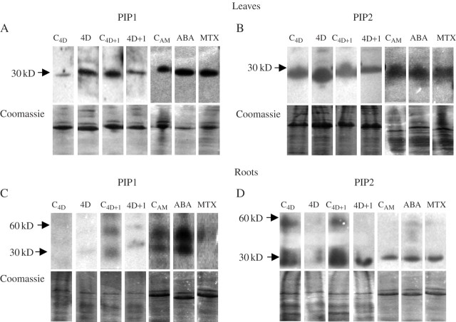
Fig. 6.
Western blots using antibodies against PIP1 (A and C) and PIP2 (B and D) proteins in leaves (A and B) and in roots (C and D) of common bean plants subjected to drought for 4 d (4D) and 1 d after re-watering (4D + 1), and the corresponding control plants (C4D and C4D+1), leaves sprayed 24 h earlier with 100 μm ABA or 200 μm MTX, and unsprayed control plants (CAM). Blots were repeated three times with different sets of plants; representative blots are shown. The corresponding Coomassie-stained gel shows the protein loaded in each lane.

