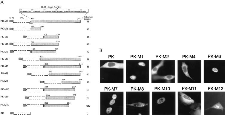
Figure 3.
Delineation of the HuR nuclear localization signal. (A) Schematic diagram and summary of the intracellular localization of PK fusion proteins containing HuR fragments M1 to M12. (B) NIH 3T3 cells were transiently transfected with myc-tagged PK expression vectors containing HuR fragments and processed as described in Fig. 1B, except for use of anti-myc antibody 9E10. myc-tagged PK was transfected as a control.

