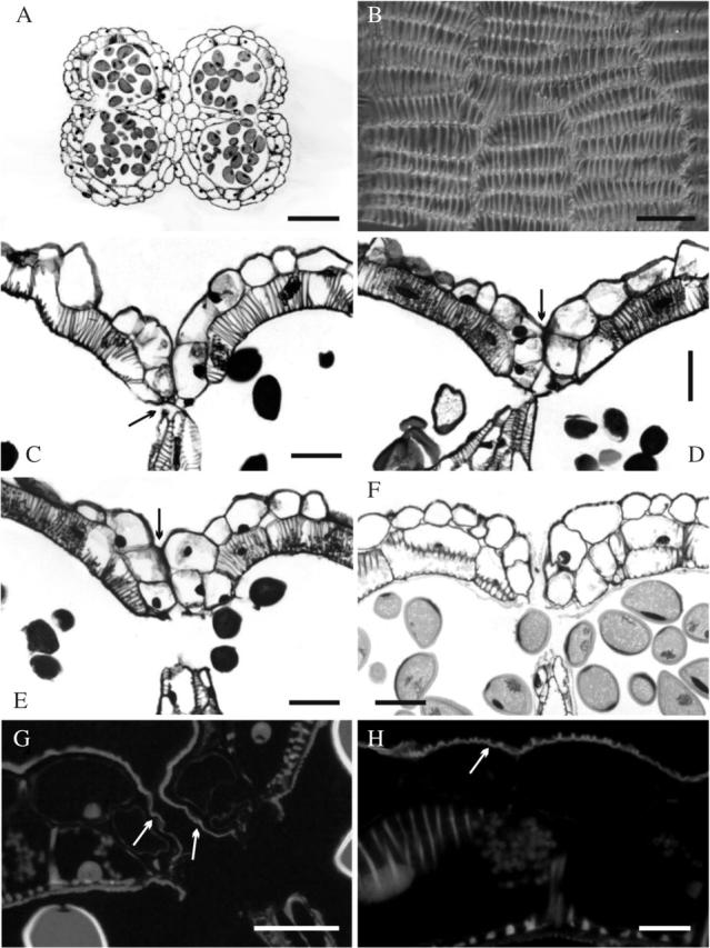
Fig. 2.

Histological features of Allium triquetrum anther. (A and C–H) Cross sections: (A) the anther has dorsiventral symmetry—note wall thickenings in the endothecium; (B) inner tangential view of the endothecium, showing tangentially elongated cells and inner bars of helical and U-shaped thickenings (some branched); (C) stomium detail before breakage in a flower bud immediately before anthesis, showing two small cells (arrow) and the septum breaking away; (D–F) successive stages during the first day of flower anthesis; (D) stomium opening—note shrinking septum and epidermal cells lining the stomium still adhering (arrow); (E) open stomium but anther still closed by epidermal cells adhering to each side of the stomium (arrow); (F) opening of the anther (i.e. open stomium, separated walls); (G) opening of the anther (i.e. open stomium, separated walls) under UV light; note smooth cuticle (arrows) lining the stomium; (H) detail of the middle part of the anther wall under UV light, showing ridged cuticle, which is common on the epidermis. Stains: A and C–F, Toluidine Blue; G and H, Auramine O. Scale bars: A = 200 µm; B–G = 50 µm; (H) 15 µm.
