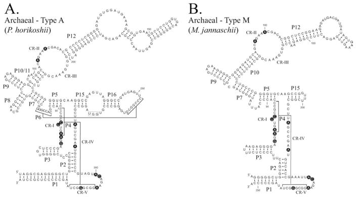
FIGURE 3.
Comparision of Type A and Type M archaeal RNase P RNAs. The secondary structures of RNase P RNAs derived by phylogenetic comparative sequence analysis are shown (Haas et al., 1996). (A). The secondary structure of the Pyrococcus horikoshii OT3 RNase P RNA (Type A). (B). The secondary structure of the Methanococcus jannaschii RNase P RNA (Type M). Structures are represented in a similar manner to Figure 2. Nucleotides conserved in all known RNase P RNAs are shown in bold background.

