
Figure 3.
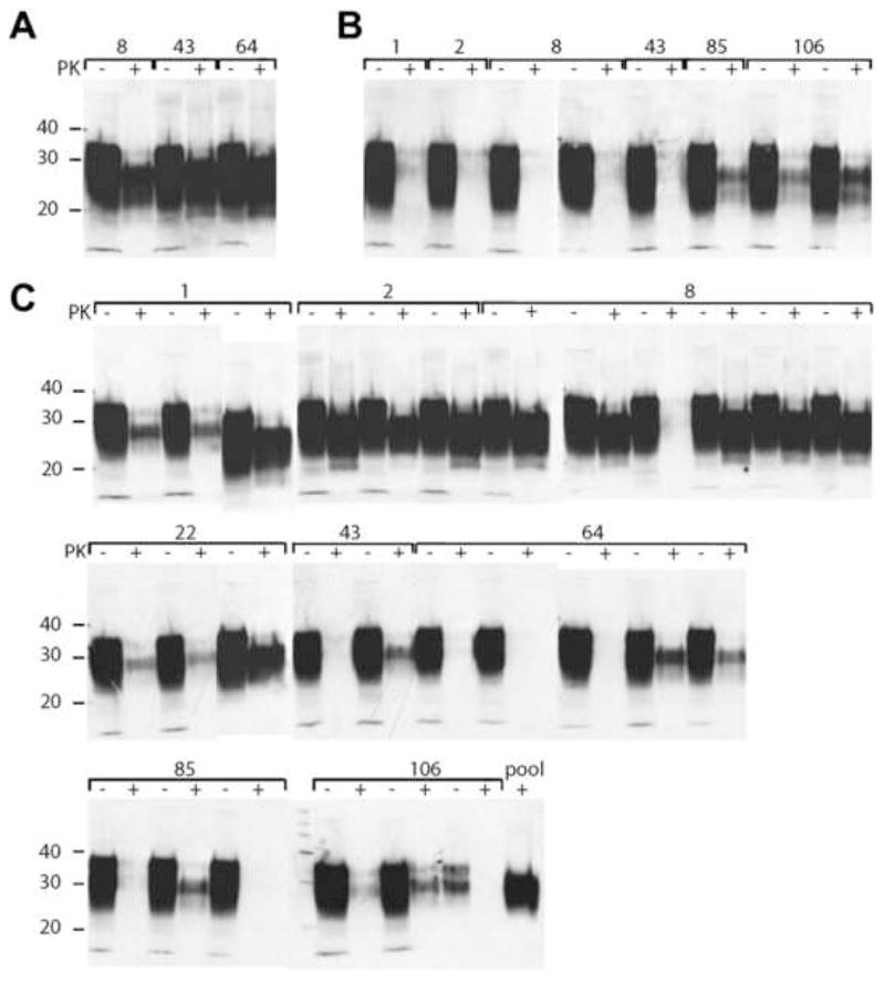
Western blot analysis of brain homogenates of Tg7 mice inoculated intracerebrally with irradiated fecal homogenates. Inocula were feces collected at 1, 2, 8, 22, 43, 64, 85, and 106 days, as indicated, from intracerebrally inoculated Syrian hamsters (A) intraperitoneally inoculated Syrian hamsters (B), and orally infected Syrian hamsters (C). PTA pellets of PK-treated brain homogenates (+) were resuspended in 150 μl of 2× sodium dodecyl sulfate (SDS) sample buffer [57]. Undigested brain homogenates (−) were diluted to 1.2% (w/v) in 1 SDS sample buffer. Equal volumes of undigested and digested samples were boiled for 5 min prior to electrophoresis. SDS gel electrophoresis and Western blotting were performed as previously described [2]. PrP was detected with the recFab HuM-P and developed with the enhanced chemiluminescent detection system (Amersham Biosciences).