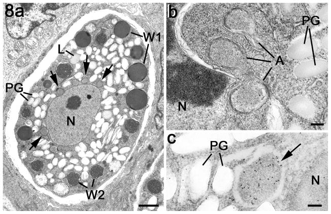
Fig. 8.
Electron micrographs of apicoplasts within the macrogametocyte. (a) Mature macrogametocyte showing the centrally located nucleus (N) with the cytoplasm containing peripherally located wall forming bodies type 1 (W1), wall forming bodies type 2 (W2), numerous lipid droplets (L) and polysaccharide granules (PG). Note the membrane-bound structures representing the apicoplasts around the nucleus (arrows). Scale bar = 2 μm. (b) Detail from the periphery of a nucleus (N) showing a number of profiles of the apicoplast. PG – polysaccharide granules. Scale bar = 100 nm. (c) Immunoelectron microscopy of a section through a macrogametocyte labelled with anti-EtENR showing numerous gold particles over the apicoplast (arrow) located between the nucleus (N) and the polysaccharide granules (PG). Scale bar = 100 nm.

