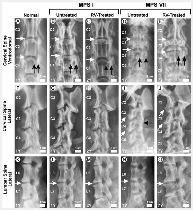
Figure 1. Radiographs of vertebrae.
Radiographs from male dogs were obtained at the ages in years (Y) shown in the lower left corner. The genotype and treatment status are indicated above the panels. RV-treated dogs received neonatal IV injection of an RV expressing the appropriate gene. Examples of RV-treated MPS I dogs are from I-171 in panels C and H and I-172 in panel M, while all examples of MPS VII dogs are from M1328. The scale bar shown in each image is 1 cm, and the cranial aspect is at the top and the caudal aspect at the bottom. A–E. Ventrodorsal view of the cervical spine. The second cervical vertebra (C2), C3, and C4 are indicated. The horizontal white arrows indicate intervertebral spaces. Black vertical arrows indicate the medial and lateral borders of the pedicle, and the space between the arrows was used to assess width. F–J. Lateral view of the cervical spine. Slanted white arrows indicate caudoventral vertebral body beaking. The black horizontal arrow indicates fusion of the articular facet joint. K–O. Lateral lumbar spine. The sixth lumbar vertebra (L6) and L7 are indicated. Horizontal white arrows indicate intervertebral spaces.

