
Figure 4. Induction of Trim21 by NDV infection, TLR and IFN stimulation.
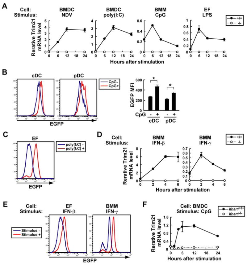
A, Trim21+/+ and Trim21−/− BMDCs, IFN-γ-primed BMMs, and EFs were infected with NDV or stimulated with TLR ligands, poly(I:C), CpG or LPS, and Trim21 mRNA was quantified by qPCR. Data are representatives of at least three independent experiments. B, EGFP MFI was measured for cDC (CD11c+B220−) and pDC (CD11c+B220+) harvested from Trim21−/− BMDCs with or without CpG stimulation for 24 h. Values are the means of three experiments ± SEM. *, p < 0.05. C, EGFP MFI was measured for Trim21−/− EFs cultured with or without poly(I:C) for 24 h. D, Unprimed Trim21+/+ and Trim21−/− BMMs were stimulated with IFNs, and Trim21 mRNA was quantified by qPCR. Data are representatives of at least three independent experiments. E, EGFP MFI was measured for Trim21−/− EFs or unprimed BMMs stimulated with or without IFN-β or IFN-γ, respectively, for 24 h. F, Ifnar1+/+ and Ifnar1−/− BMDCs were stimulated with CpG, and Trim21 mRNA was quantified by qPCR.