Figure 3.
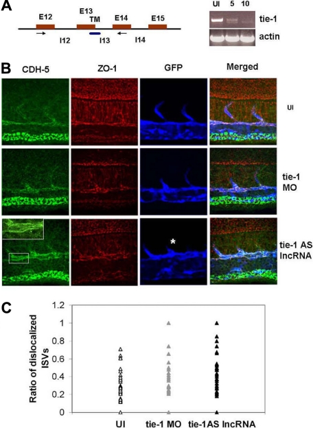
The phenotype of zebrafish tie-1AS lncRNA overexpression. (A) The MO targeting splice-site is complementary to the 13th exon-intron boundary. RT-PCR was performed to confirm MO-targeting effects. (B) Immunostaining of Tg(flk: EGFP) embryos injected with tie-1AS (150 pg) or tie-1 MO (5-10 ng) at the 1-cell stage and fixed at 24 hpf. Staining was performed using CDH5, ZO-1, and anti-GFP antibodies. Most tie-1AS lncRNA-injected embryos showed an asymmetric distribution of Cdh-5 staining on endothelial cell membrane in vivo as shown in the enlarged inset. ISVs (white asterisk) also show a truncated phenotype. Details of image capture are available in supplemental Methods. (C) Quantitation of the phenotype was performed as described in supplemental Methods, and the ratio of length of ISVs showing phenotype to an ISV with normal CDH-5 distribution is plotted. In general, the trend in tie-1AS lncRNA and tie-1 MO-injected embryos shows higher ratios indicating more ISVs with asymmetric CDH-5 distribution per embryo.

