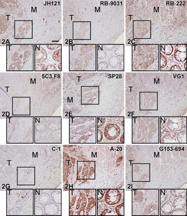
Figure 2.
IHC staining results in nine VEGF antibodies on paraffin colon adenocarcinoma tissue sections. Boxed area of the tumor part is magnified in the left inset (T); in the right inset (N) is a detail of the adjacent normal colon. M, muscularis propria. (D,G) Note negative staining of tumor (T) and normal epithelium (N). All other VEGF antibodies showed variable staining patterns. Bar = 0.1 mm.

