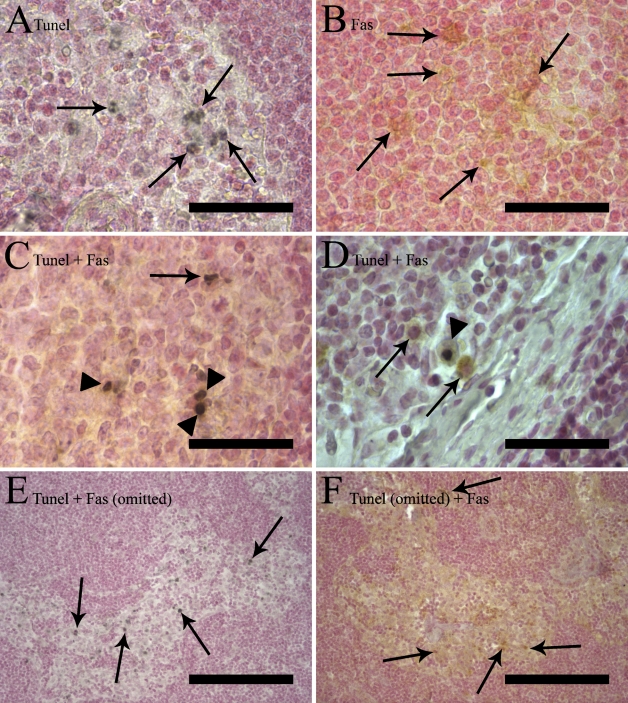
Figure 1.
Five different combinations of terminal deoxyribonucleotidyl transferase–mediated dUTP nick end labeling (TUNEL) and immunostaining of the same sample. Counterstaining was performed with Nuclear Fast Red. (A,B) Single staining. (A) A single TUNEL staining labeling apoptotic nuclei in a germinal center black diaminobenzidine (DAB-black) chromogen. Arrows indicate single TUNEL stained cells. (B) Fas single staining with brown label (DAB chromogen) in the cytoplasm (arrows), leaving the nucleus unstained. (C,D) Double staining with TUNEL and Fas showing many double-stained cells with a black nucleus and brown cytoplasm (arrowheads). There are also single-positive cells (arrows) in both images, TUNEL-positive in C and Fas-positive in D (arrows). (E,F) Control staining in which either the primary antibody (anti-Fas) or terminal deoxynuleotidyl transferase (TdT) enzyme has been omitted. Arrows indicate single-positive cells. (E) After omission of the Fas primary antibody in a double staining (sinus), there is no brown coloration of the immunoreaction at all in the section, whereas the TUNEL staining still shows a staining pattern similar to that seen in A, C, and D. (F) After omission of the TdT enzyme in a double staining (sinus), only Fas-positive brown cells are visible. Bars: A–D = 50 μm; E–F = 200 μm.

