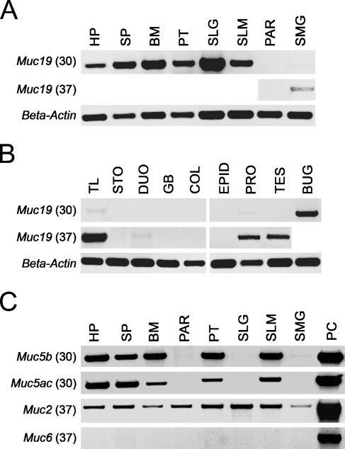
Figure 1.
Expression of Muc19 transcripts in mouse tissues. (A) Upper panel: RT-PCR products using 50 ng of random primed cDNA at 30 cycles from major and minor salivary glands. HP, hard palate; SP, soft palate; BM, buccal mucosa; PT, posterior tongue; SLG, sublingual gland; SLM, sublingual mucosa; PAR, parotid gland; SMG, submandibular gland. Middle panel: tissues with no products at 30 cycles were re-tested at 37 cycles. Lower panel: β-actin–positive controls for each tissue sample (30 cycles, 349-bp product). (B) RT-PCR results for non-salivary tissues (upper and middle panels) as described for A. TL, tracheolarynx; STO, stomach mucosa; DUO, duodenum; GB, gall bladder; COL, colon; EPID, epididymis; PRO, prostate; TES, testes; BUG, bulbourethral gland. (C) Expression of transcripts for the gel-forming mucins Muc5b, Muc5ac, Muc2, and Muc6 in salivary tissues. Shown are RT-PCR products using 50 ng of cDNA at either 30 cycles (Muc5b and Muc5ac) or 37 cycles (Muc2 and Muc6). Abbreviations are as given in A. PC, positive control tissue used for each mucin, tracheolarynx (Muc5b), stomach mucosa (Muc5ac and Muc6), and small intestine (Muc2).

