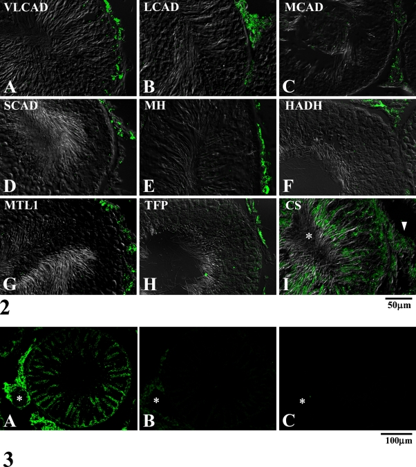
Figure 3.
Immunofluorescent micrographs of adult rat testis observed at low-power magnification for showing control stainings. Tissue sections were stained with the antibody for a mitochondrial fatty acid β-oxidation enzyme, MTL1 (A), the same antibody preabsorbed with purified enzyme of MTL1 (B), and IgG fraction of non-immune serum (C). Asterisk, small artery in the interstitial tissue. Note that the staining of Leydig cells in the interstitial tissue is relatively more intense than that of the seminiferous epithelium. Note also that preabsorption of the antibody with MTL1 enzyme markedly diminished the staining.

