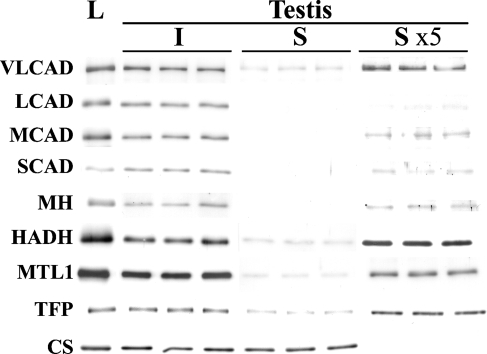
Figure 5.
Immunoblot analysis of the interstitial tissues and seminiferous tubules of the testis. The separated preparations of the interstitial tissues (I) and the seminiferous tubules (S) with 1 μg of protein were analyzed with pooled liver from three rats (L) with 1 μg of protein. The lanes marked S ×5 show blots with five times the amount of protein (5 μg). Note that all of the mitochondrial fatty acid β-oxidation enzymes are abundantly present in the interstitial tissue, whereas the signals in the seminiferous tubule are very faint, when compared with the liver tissues. In contrast, CS is present in both the interstitial tissue and the seminiferous tubule in amounts similar to those in liver tissue. (See Figure 2 legend for abbreviations.)

