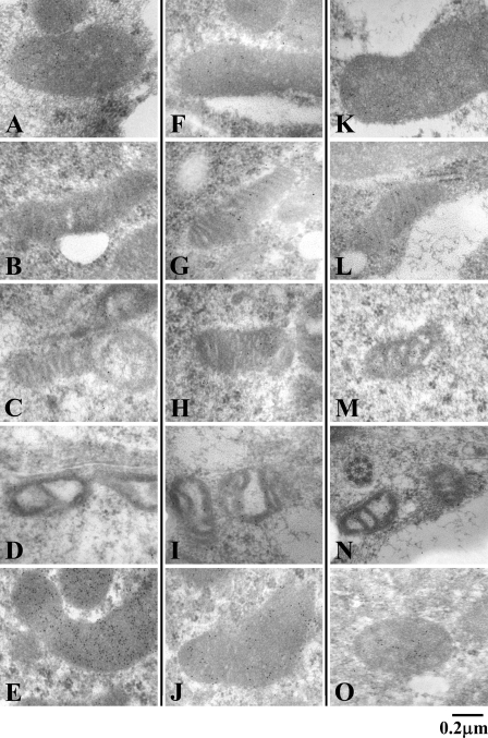
Figure 8.
Immunoelectron micrographs of mitochondria in the cells of the seminiferous epithelium at stage V–VI. Sertoli cell (A,F,K), type B spermatogonium (B,G,L), primary spermatocyte (C,H,M), spermatid (D,I,N), and Leydig cell (E,J,O), stained for MTL1 (A–E), TFP (F–J), and CS (K–O). Note that mitochondria of Sertoli cells and Leydig cells are strongly positive for the staining for MTL1, TFP, and CS; in contrast, the staining intensity of spermatogenic cells varies among the three enzymes.

