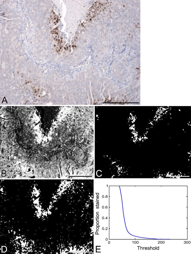
Figure 2.
(A) Image of a typical immunohistochemistry (IHC)-stained slide. Background staining is generally a light blue with this technique, and represents cells that do not express the antigen of interest. The brown stain is the product of a colorimetric assay that is of standard use in clinical IHC measurements, and represents cells that are positive for the antigen of interest. (B) Image representing the intensity of brown staining for the slide in A. Brighter pixels indicate stronger staining, dark pixels indicate no staining. (C) Thresholded version of the intensity image: pixels with intensity above 100 are identified as white; the remainder are black. (D) Another thresholded version of the intensity image: pixels with intensity above 70 are identified as white; the remainder are black. (E) Curve showing the percent area stained as a function of the threshold. Bar = 200 μm.

