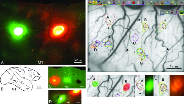
Figure 1.
Photomicrographs (M1, M2, M3) of injection sites, and alignment (for M1) with optical-imaging patterns. (A) Fluorescence photomicrograph of the 2 injections (CTB-Alexa488, at left; CTB-Alexa555 at right) in M1. Note the clear separation of both injection cores and halo. The intense injection cores have saturated the optics and appear almost white. (B) Schematic brain diagram to show the location of the 2 injections in M1. Dashed line anterior to PMTS indicates the border between the tangentially sectioned block (containing the injection sites) and the larger, coronally sectioned posterior block. (C, D) Photomicrographs of the 3 injections in M2 and of 2 of the 3 injections in M3, at slightly lower magnification (and see Figs 3 and 7 for schematic brain diagrams). (E) Photograph of the brain surface (M1), seen through the implanted chamber. Note surface blood vessels, which were used as reference landmarks in the physiological experiments and guides for the anatomical injections. Eight regions (A–H) corresponding to surface darkening during the optical-imaging sessions are indicated. Colored outlines delineate domains responsive to the object stimuli shown at the top (and coded by a comparable color bar at the bottom of the image). Small green dots represent electrode penetrations. Large red and green dots in spots (A), (B) denote 2 injection sites. Purple dots (indicated by arrowheads, left and right of spot F) mark the position of DiI. Below left: The 2 anatomical injections are overlain (in solid green and solid red) on the 2 targeted (A, B) activity spots. Below right: Two pairs of images, showing activity spots (A) and (B) and the tracer injections, photographed in vivo, just subsequent to injection (see Methods). Arrows point to the same blood vessels as in (E). Scale bars in (C) and (D) = 500 μm. AMTS = anterior middle temporal sulcus; CS = central sulcus; IAS = inferior arcuate sulcus; LS = lateral sulcus; LuS = lunate sulcus; PMTS = posterior middle temporal sulcus; PS = principal sulcus; SAS = superior arcuate sulcus.

