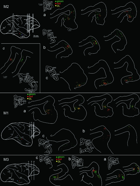
Figure 7.
Drawings of representative coronal sections showing the distribution of retrogradely labeled neurons in the PFC (see the 3 repeated color codes for population scoring). Only for M2, labeling in the intraparietal cortex is also shown (level d). In all 3 brains, the prefrontal labeling was located at 3 rostrocaudal levels (a–c). For each level we choose from 1 to 4 representative sections (200 μm apart). Small section drawings are offset to the left for orientation, and the zone corresponding to the higher magnification chartings is indicated by the dashed boxes. The dorsolateral view of the injected hemispheres is repeated at the left, for further orientation. Silver neurons, which were very few in number, are not shown (but see Table 3). IPD = infraprincipal dimple; LOS = lateral orbital sulcus. Other abbreviations as in Figure 1.

