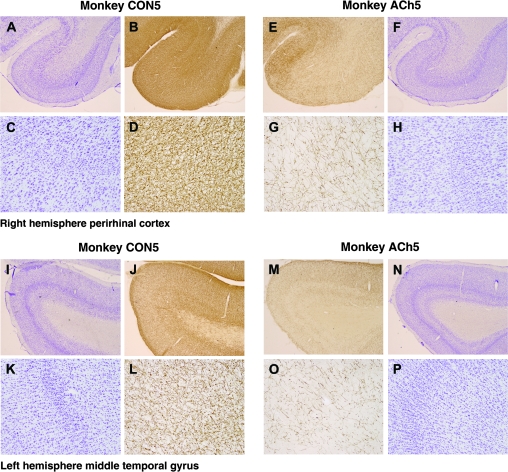
Figure 6.
Examples of low- and high-magnification cresyl violet– and AChE-stained sections from monkey CON5, who received injections of saline into inferior temporal cortex, and from monkey ACh5, who received injections of saporin. Photographs (A–D) and (I–L) show the perirhinal cortex and MTG, respectively, of control monkey CON5. Photographs (E–H) and (M–P) show the perirhinal cortex and MTG, respectively, of cholinergic lesioned monkey ACh5. In all monkeys, the toxin was effective evidenced by the loss of AChE-postive fibers, and this is clearly seen at higher magnifications (sections G and O). Cresyl violet sections, adjacent to those processed for AChE, showed no gross abnormalities.

