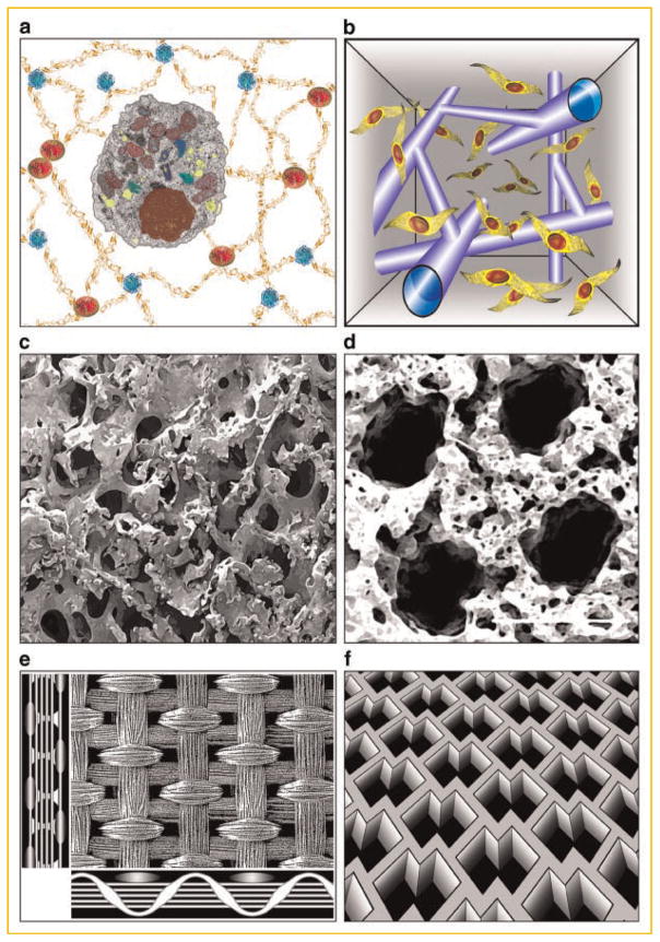
Fig. 3.
Structural signals provided through scaffold design. a: Hydrogels with tunable molecular, mechanical, and degradation properties. b: Post-gelation modification of hydrogel scaffold by laser light enables geometrically precise degradation of hydrogel, to form channels for cell migration [Kloxin et al., 2009]. c: Mechanically strong, highly porous, mineralized silk scaffold for bone tissue engineering [Wang et al., 2006]. d: Soft, highly porous, channeled elastomer scaffold for engineering vascularized cardiac muscle [Radisic et al., 2006]. e: Knitted matt-gel composite scaffold with structural and mechanical anisotropy for cartilage tissue engineering [Moutos et al., 2007]. f: Accordion-like elastomer scaffold with structural and mechanical anisotropy designed for cardiac tissue engineering [Engelmayr et al., 2008].

