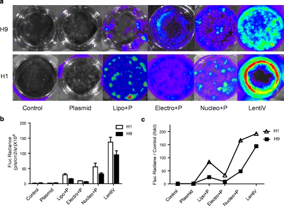
Fig. 2.

Transfection and transduction of H9 and H1 hES cells in vitro with each of the transfection protocols. (a) H9 and H1 cells were seeded onto feeder-cell free Matrigel-coated six-well plates after transfection with plasmid alone (Plasmid), lipofection (Lipo+P), electroporation (Electro+P), nucleofection (Nucleo+P), and transduction with lentivirus (LentiV). (b) The cells were examined using bioluminescence imaging of Fluc activities after transfection. The quantitative measurements are shown 24 h after gene delivery.
