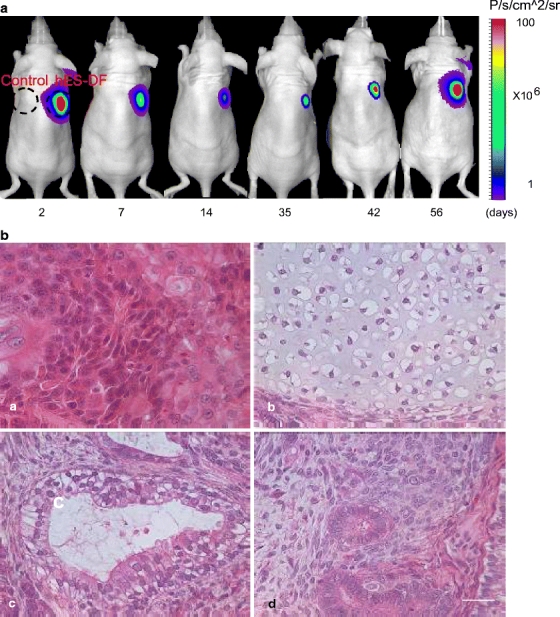
Fig. 5.

Optical bioluminescence imaging of stably expressed hES cells in vivo over time, with demonstration of proliferation and teratoma formation. The pUb-eGFP-Fluc-SV40-Puro construct was stably expressed in H9 cells by transduction with replication-incompetent lentivirus. Stable colonies (hES-DF) were selected by drug selection over 2 weeks and then injected subcutaneously into right shoulder of nude mice. Control untransduced H9 hES cells were injected into left shoulder. (a) In vivo bioluminescence imaging of the hES-DF cells on days 2, 7, 14, 35, 42, and 56. (b) Histology of the H9 hES cells stably expressing eGFP-Fluc in nude mice were examined through 56 days. Teratoma formation was demonstrated by histology at week 8 weeks after subcutaneous injection: (A) rosette consistent with neuroectodermal differentiation (ectoderm), (B) cartilage formation (mesoderm), (C) respiratory epithelium with ciliated columnar, and (D) mucin-producing goblet cells (endoderm) surrounded with mesenchymal cells (mesoderm). Scale bar = 50 µm.
