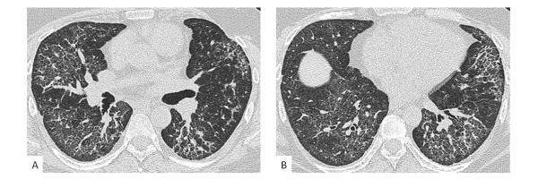
Figure 3.
High resolution thoracic CT three years after diagnosis. High resolution CT sections at the level of a) the hilae and b) the lung bases demonstrate resolution of the previously noted consolidation and a reduction in ground glass attenuation. However evidence of fibrosis persists with bilateral reticular change and traction bronchiectasis. The dome of the right hemidiaphragm is visible in the image of the lung bases (b).

