
Figure 3.
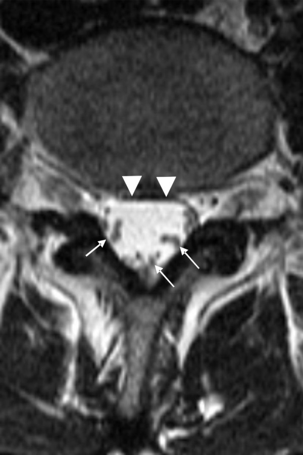
Magnetic resonance images of a normal lumbar spine. Cross-sectional magnetic resonance images of a normal lumbar spine with dural sac cross-sectional area of 180 mm2 and LEL 0 (according to Borré). On the T2-weighted image, the cerebrospinal fluid appears nearly white and the nerve roots are more easily seen in the large cerebrospinal volume than on a T1-weighted image. The nerve roots are free to move away in the cerebrospinal fluid space when a needle is inserted into the dural sac.