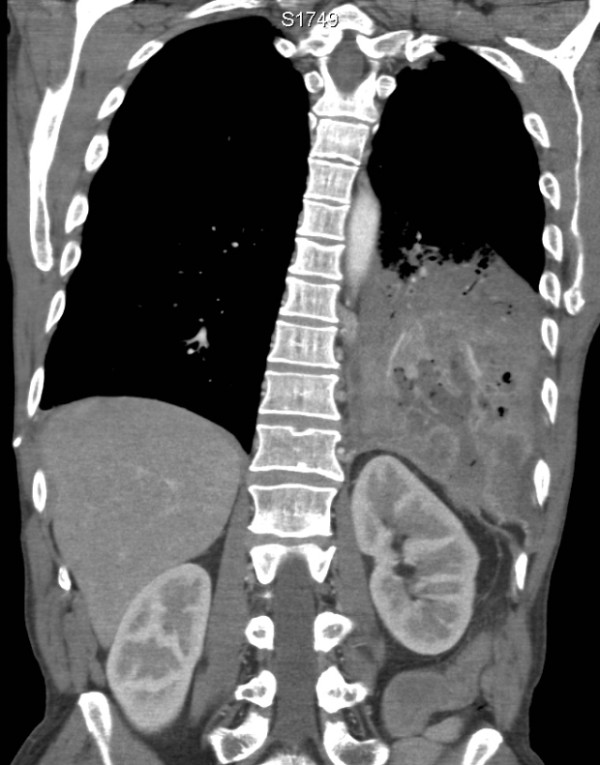
Figure 2.

Coronal CT image shows the cavitating left lower lobe pneumonia. The fluid- and gas-containing collection transgresses the diaphragm and enters the retroperitoneal space. Although not shown here, this collection is contiguous with an abnormally thickened splenic flexure.
