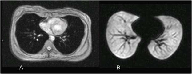
Figure 3.
Normal subject. (A) Conventional axial 1H MR image of the chest showing the soft tissues of the chest. The airspaces of the lung show as dark due to the low water content. (B) Corresponding 3He MR image obtained immediately after inhalation of the hyperpolarized helium-3 gas shows homogeneous, bright appearance of the gas-filled airspaces. The dark linear structures within the lungs represent the pulmonary vessels. Reprinted with permission from Chest 2006;130:1055–62.

