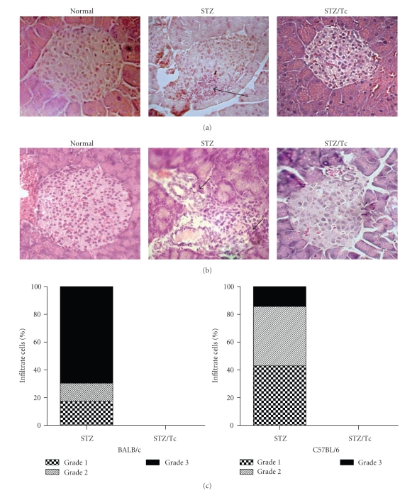
Figure 3.
MLDS-mediated insulitis in BALB/c mice. All mice received MLDS for 5 consecutive days and were sacrificed 6 weeks later to harvest tissues for histopathology. (a) H&E stained BALB/c mouse pancreas sections: normal islet, STZ (uninfected and MLDS-treated), STZ/Tc (T. crassiceps-infected and MLDS-treated). (b) H&E stained C57BL/6 mouse pancreas sections. (c) Score of cell infiltrates in BALB/c and C57BL/6 islets. Note the lack of leukocyte infiltrates in T. crassiceps-infected mice. Arrows indicate infiltration. Magnification ×400.

