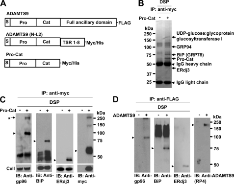
FIGURE 1.
Identification of molecules that interact with ADAMTS9 Pro-Cat. A, domain structure of the previously published ADAMTS9 constructs used in this analysis. TSR, thrombospondin type 1 repeats. B, HEK293 F cells stably expressing ADAMTS9 Pro-Cat were cross-linked with membrane-permeable, thiol-cleavable DSP, and immunoprecipitated with polyclonal anti-Myc antibody as described. The eluted samples were separated by NuPAGE (4–12% gradient gel), and the indicated bands (arrows) were cut out and identified by LC/MS/MS. C, following cross-linking and immunoprecipitation with polyclonal anti-Myc, immunoprecipitates were analyzed by Western blotting with monoclonal anti-gp96 antibody, monoclonal anti-BiP antibody, polyclonal goat anti-ERdj3 antibody, or monoclonal anti-Myc antibody as indicated. The lower panel shows total cellular expression of gp96, BiP, ERdj3, and Pro-Cat in nontransfected (−) or transfected (+) cells. A protein complex (*) is recognized by both anti-gp96 and anti-Myc, but not by anti-BiP and anti-ERdj3. D, full-length ADAMTS9 (panel A) was affinity-isolated using anti-FLAG® M2 Affinity Gel, and the eluted proteins were analyzed by Western blotting with the same antibodies as used above. Anti-RP4 antibody was used to detect ADAMTS9. Arrowheads indicate the individual protein bands corresponding to (from left to right), gp96, BiP, ERdj3, and ADAMTS9.

