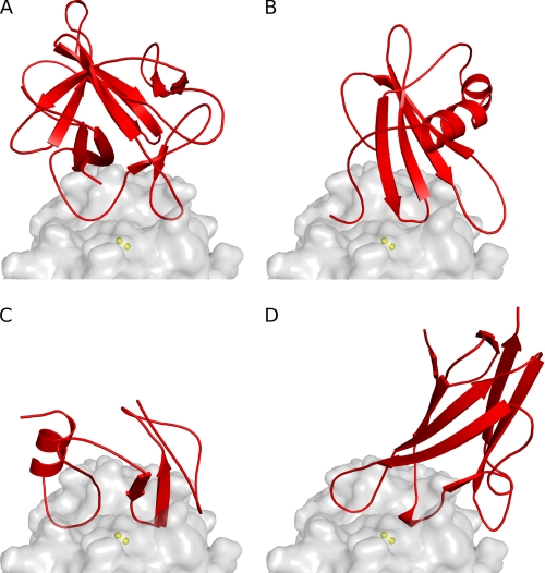
FIGURE 5.
Comparison of the binding modes of inhibitors. The fold of inhibitors is shown in red (A, clitocypin; B, stefin A; C, p41 fragment; D, chagasin), whereas the endopeptidase cathepsin V is presented as a gray surface. The surface of the catalytic cysteine is shown in yellow. All inhibitors have different folds, yet for docking to the active site of papain-like cysteine proteases they utilize loops that fill the active site along its whole length and occlude the reactive site cysteine residue by two loops.

