FIGURE 2.
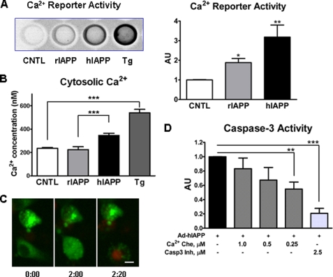
Overexpression of hIAPP in INS 832/13 cells leads to elevation of cytosolic Ca2+, NFAT-GFP nuclear translocation, and cell death. INS 832/13 cells were transduced with rIAPP or hIAPP adenovirus at 400 m.o.i. A, cytosolic Ca2+ was measured using NFAT-SEAP gene reporter system 12 h post-transduction with adenovirus (see “Experimental Procedures” for details). CNTL, control. B, Ca2+ concentrations were measured ratiometrically 6 h post-transduction by monitoring more than 100 individual cells loading with 5 μm Fura-2/AM. Positive control was 1 μm of Ca2+-ATPase inhibitor thapsigargin (Tg). C, INS 832/13 cells were transfected with plasmid DNA expressing NFAT-GFP for 12 h and then transduced with Ad-hIAPP at 400 m.o.i. for 8 h. Then laser confocal microscopy was performed to follow individual cells with elevated cytosolic Ca2+ for 2 h in the presence of 500 ng/ml PI. The cell at the bottom showed cytosolic NFAT-GFP distribution at the beginning of observation. Two hours later, NFAT-GFP in this cell translocated to the nucleus, and then 20 min later the nucleus incorporated propidium iodide indicating cell death. The scale bar is 5 μm. D, INS 832/13 cells were transduced with hIAPP adenovirus (Ad) for 6 h, and then three doses of Ca2+ chelator (Ca2+ Che) BAPTA-AM were applied for 48 h. Caspase-3 (Casp-3) inhibitor was used to show the assay specificity and as a positive control. Data are the mean ± S.E. from four (A) and five experiments (D); *, p < 0.05; **, p < 0.001; ***, p < 0.0001. AU, arbitrary unit.

