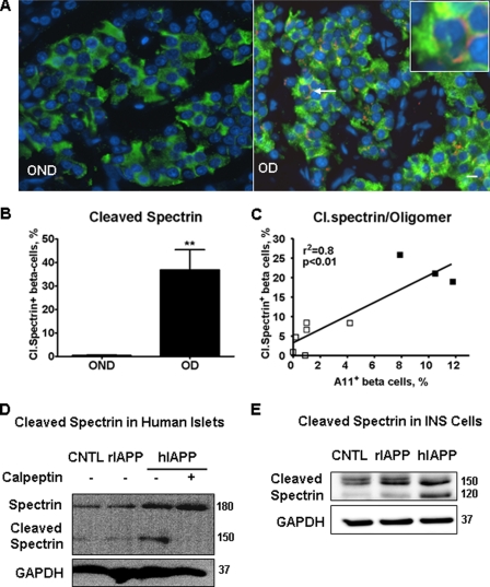
FIGURE 6.
Cleavage of calpain substrate spectrin was detected in beta cells of paraffin-embedded pancreatic tissues from humans with T2DM. A, immunofluorescent images of pancreatic sections stained with anti-cleaved spectrin (red) and anti-insulin antibodies (green). Nuclei were visualized by DAPI (blue). Beta cell with cleaved (Cl.) spectrin was indicated by an arrow (inset shows beta cell with cleaved spectrin in the periphery). B, quantification of beta cells positive for cleaved spectrin in tissue sections from age- and BMI-matched obese nondiabetic (OND, n = 7) and from obese diabetic subjects (OD, n = 7). C, there was a positive correlation between frequency of beta cells with cleaved spectrin and frequency of beta cells positive for toxic oligomers detected by immunofluorescent labeling with oligomer-specific antibody A11 in surgically obtained human pancreas. These tissue samples were previously used to report the increased frequency of toxic IAPP oligomers in beta cells of individuals with T2DM compared with nondiabetic controls (15). □, nondiabetic; ■, T2DM. D, overexpression of hIAPP in human islets induced spectrin cleavage that was blocked by calpain inhibitor calpeptin. Islets were transduced with hIAPP or rIAPP as control at m.o.i. 400 for 72 h. CNTL, control; GAPDH, glyceraldehyde-3-phosphate dehydrogenase. E, cleavage of spectrin was induced by overexpression of hIAPP in INS 832/13 cells. Cells were transduced with hIAPP or rIAPP as control at 400 m.o.i. for 48 h. Data are the mean ± S.E.; **, p < 0.01.

