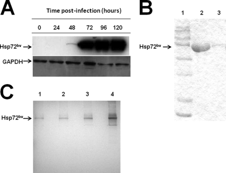
FIGURE 3.
Expression of recombinant human Hsp72bv in Sf9 insect cells. A, Sf9 insect cells (2 × 10⁶ cells/ml) were infected with recombinant baculovirus virus containing the hsp72 gene. Samples were collected every 24 h post-infection and examined for the expression of Hsp72 by Western blot analysis. Briefly, membranes were probed with mouse anti-Hsp72 monoclonal antibody (top panel) or anti-GAPDH (loading control; bottom panel) followed by incubation with peroxidase-conjugated goat anti-mouse IgG secondary antibody. Lane 1, 0 h; lane 2, 24 h; lane 3, 48 h, lane 4, 72 h; lane 5, 96 h; and lane 6, 120 h. In a separate experiment 96 h post-infection, cells were collected, and clear cell lysate was applied to a Ni-NTA His·Bind resin column. Purified protein Hsp72bv was collected and desalted using Centricon Ultracel YM-50. Hsp72bv protein was analyzed using SDS-PAGE followed by either Coomassie blue staining (B); lane 1, protein maker; lane 2, 30 μg Hsp72bv; lane 3, 1.5 μg Hsp72bv or Silver staining (C); lane 1, 50 ng Hsp72bv; lane 2, 100 ng Hsp72bv; lane 3, 200 ng Hsp72bv; lane 4, 500 ng Hsp72bv. The data is a representative experiment from three independently performed experiments with similar results.

