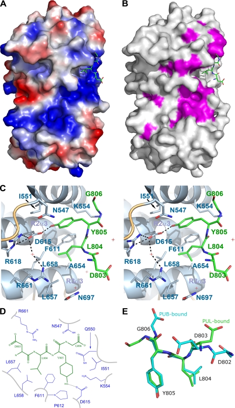
FIGURE 2.
Co-structure of the PUL domain with the C-terminal p97 peptide. A, electrostatic surface representation was generated with a gradient from −10 (red) to 10 (blue) kT/e of the PUL domain only. The bound p97 peptide is show in green stick format. B, surface representation of the PUL domain is shown in light gray with conserved residues in purple. C, stereoscopic view of the terminal p97 peptide residues LYG806 bound to the PUL domain shown with helices in schematic format and labeled. PUL residues within proximity are shown in stick format and labeled. The hydrogen bond is shown as a black dashed line; water molecules are shown as red cross-hairs. D, schematic representation of p97 (black) interactions with PLAA (blue). E, three-dimensional alignment of the PLAA (green) and PNGase (cyan)-bound p97 peptides.

