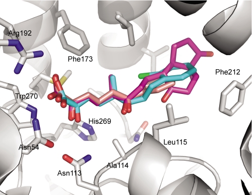
FIGURE 5.
Overlay of the MCP substrates HOPDA, HOPODA, and DSHA in complex with S114A. The protein (white) is shown in with the side chains involved in ligand binding shown as sticks. The ligands are represented as sticks and are colored pink (HOPDA), cyan (HOPODA), and purple (DSHA). In all structures the nitrogen, oxygen, and sulfur atoms colored blue, red, and yellow, respectively.

