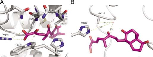
FIGURE 6.
Interaction of DSHA with S114A. A, the structure of S114A (white) in complex with DSHA (purple) is shown in cartoon representations with the amino acid side chains involved in binding and DSHA displayed in stick representation. The interactions between S114A and the C-2 oxo and C-1 carboxylic acid are shown by yellow dashes, whereas the interactions between C-6 oxo and S114A are shown by red dashes. B, the structure of S114A (white) in complex with DSHA (purple). The water molecule (HOH89) is shown by a red star. All of the distances are in angstroms.

