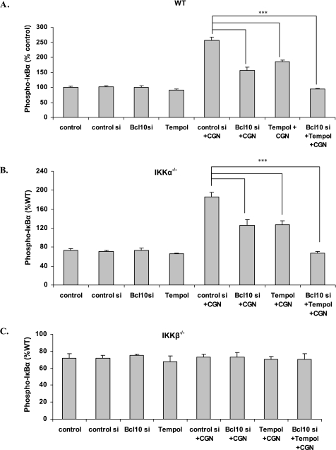
FIGURE 3.
Differential increases in phospho-IκBα in the MEF IKKα−/− and β−/− cells. A, in the MEF WT cells, the CGN-induced increases in phospho-IκBα were reduced by Tempol and by BCL10 silencing (p < 0.001). B, results were similar in the IKKα−/− cells (p < 0.001). C, in contrast, in the IKKβ−/− cells, phospho-IκBα did not increase following CGN exposure.

