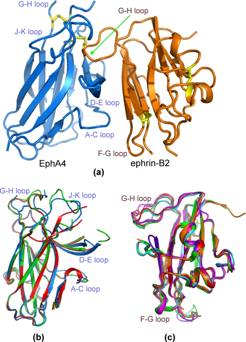
FIGURE 1.
Crystal structure of the EphA4-ephrin-B2 complex. a, overall structure of the EphA4-ephrin-B2 complex. Cysteines involved in disulfide bonds (Cys45–Cys163 and Cys80–Cys90 in EphA4 and Cys65–Cys104 and Cys92–Cys156 in ephrin-B2) are indicated in yellow. b, superimposition of the structures of EphA4 in complex with ephrin-B2 (blue) and the previously determined structures of free EphA4 molecule A (green) and molecule B (red) (7). c, superimposition of the structures of ephrin-B2 in complex with EphA4 (brown), EphB2 (purplish blue; Protein Data Bank code 1KGY), EphB4 (violet; code 2HLE), Hendra virus attachment protein (cyan; code 2VSK), and Nipah virus attachment protein (red; code 2VSM) as well as free ephrin-B2 (green; code 1IKO).

