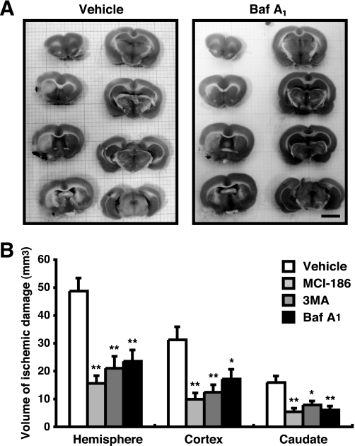
FIGURE 7.
Autophagy/lysosome inhibitors reduce volume of neural cell death. A, 3 h after MCA occlusion, ischemic damages were detected using TTC-staining. In TTC-stained slices, the infracted tissue appeared white, whereas the intact tissue was colored. Bar, 5 mm. B, the infract volume was measured and represented by the means ± S.E. *, p < 0.05; **, p < 0.01; 1-factor analysis of variance followed by Tukey-Kramer test.

