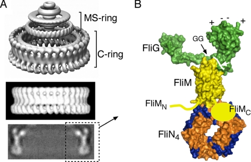
FIGURE 1.
A, images of the flagellar basal body from EM reconstructions. The upper panel shows the C-ring, MS-ring, and proximal rod. The lower, thicker part of the MS-ring is situated in the membrane; the C-ring is in the cytoplasm. The middle panel is a side view of only the C-ring, and the lower panel shows a cross-section through the C-ring. The figure has been taken from Ref. 4 with permission. B, molecular model for major portions of the C-ring based on crystal structures of major portions of the proteins and biochemical and mutational studies of subunit relationships. FliGC is on the right and contains a set of conserved charged residues (top) that interact with the stator protein MotA. FliGC is joined to the rest of the protein by a conserved Gly-Gly linker that might allow relative movement of the domains. The structure of FliMC is unknown; this domain is known to interact with FliN, but the details of this relationship are likewise unknown.

