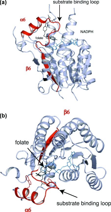
Figure 2.
PTR1 subunit architecture and position of the active site. (a) Side view of the subunit of the ternary complex with cofactor and folate. α6, β6, and the substrate binding loop are colored red. The cofactor and folate are depicted as blue and black sticks, respectively. (b) Orthogonal view to (a) in the orientation used for all other molecular images. Trp221 is represented as stick model on α6.

