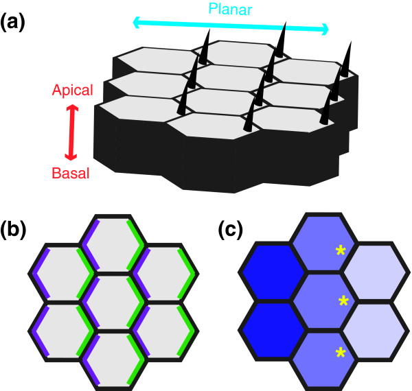
Figure 1.
Planar cell polarity. (a) Epithelial tissues display apical-basal and planar polarity. Hair structures are generated at the apical (top) surface and are absent from the basal (bottom) surface, demonstrating asymmetry along the apical-basal axis. Planar polarity is evident from the fact that the hairs are placed at the distal (right) side of each cell's apical surface and point in a distal direction. (b) Generating planar polarity through asymmetric protein localization. Schematic depicting a bird's-eye or planar view of the apical surface of the wing. The proximal (left) and distal (right) sides of each cell are defined by specific Frizzled system proteins, depicted in purple and green, respectively. (c) Generating planar polarity through protein gradients. In the Drosophila wing, the cadherin Dachsous (blue) is expressed in a decreasing gradient from proximal to distal (left to right). As a result, the cadherin Fat, which is also present in cell membranes, is predicted to be more active at the distal cell surface (yellow asterisks), where it encounters less inhibitory Dachsous protein on the adjacent cell.

