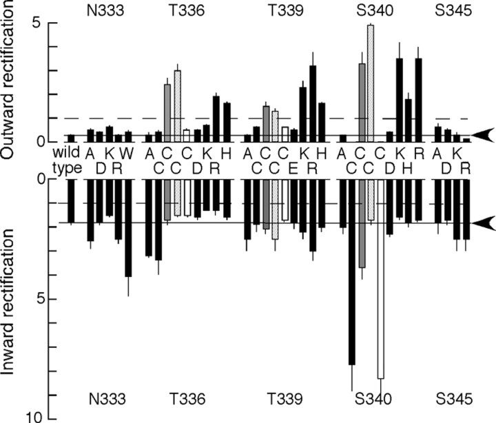
Figure 8.
Summary results for mutations causing outward rectification. At Thr336, Thr339, and Ser340, positively charged residues or positively charged MTS compounds introduced marked outward rectification. Rectification is expressed as the additional current observed at +150 mV (outward rectification, top) or −150 mV (inward rectification, bottom) compared with that which would be observed by linear extrapolation of the current–voltage relation from the range −5 to +5 mV; therefore, unity (broken line) indicates no rectification. Solid line and arrowheads indicate rectification in wild-type P2X receptors. Filled gray columns, Cysteine substitution after addition of MTSEA. Stippled columns, Cysteine substitution after addition of MTSET. Open columns, Cysteine substitution after addition of MTSES. ATP concentrations were as follows (in μm): wild-type, 3; N333A, 1; N333D, 3; N333K, 0; N333R, 0; N333W, 1; T336A, 3; T336C, 3; T336K, 0; T336R, 0; T336H, 10; T339A, 1; T339E, 3; T339C, 3; T339K, 1; T339R, 1; T339H, 3; S340A, 10; S340C, 30; S340D, 0; S340H, 0; S340K, 0; S340R, 0; S345A, 10; S345D, 10; S345K, 10; S345R, 10. Where the concentration is indicated as zero, the rectification of the “standing” current introduced by the substitution is presented.

