
Figure 9.
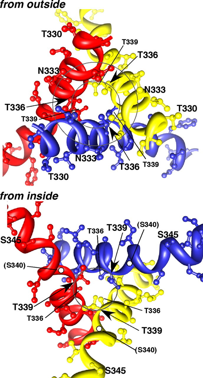
Model of three TM2 helices to illustrate positions of key residues. Viewed from the outside, the circles pass through the Cα atoms of Asn333 (outer circle) and Thr336 (inner circle) from each chain. Viewed from the inside, the circle passes through the Cα atoms of Thr339. The structure shown is a threaded model of rat P2X2 generated using a default script in Modeler9v6 from the zebrafish P2X4 receptor in the closed state (pdb 3H9V) (Kawate et al., 2009) and indicating side-chains from Asn333 to Ser340 only. The side-chain of Ser340 is not seen, but the position of Ser340 Cα is indicated (white dots). The results indicate that Ser340 is exposed in the open channel permeation pathway, which implies that the three TM2 helices twist counter-clockwise during channel opening.