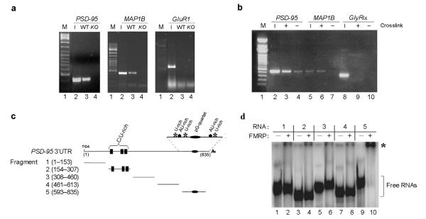
Figure 1. FMRP interacts directly with the 3′UTR of PSD–95 mRNA.
(a) Brain lysates from wildtype (WT) and FMR1 knockout mice (KO) were immunoprecipitated with FMRP antibodies. RT–PCR was performed using oligos for the PSD–95, MAP1B and GluR1 mRNAs. Input (1/5) is reported in lanes 2. Lanes not relevant to this experiment were removed between the marker and lanes 1 and 2. (b) CLIP assay. Hippocampal cell extracts were immunoprecipitated with FMRP antibodies. RT–PCR was performed using oligos for the PSD–95, MAP1B and GlyRα mRNAs. Input (1/5) is reported in lanes 2, 5, 8. (c) PSD–95 3′UTR fragments utilized in EMSA experiments. Potential functional motifs are indicated. (d) 32P radiolabelled fragments (1–5) of the PSD-95 3′UTR were incubated in the presence of FMRP (+, lanes 2, 4, 6, 8, 10). Control reactions were performed in buffer alone (−, lanes 1, 3, 5, 7, 9). RNA:protein complexes were resolved on native polyacrylamide gel. Unbound RNA fragments (]), and RNA:protein complexes (*) are indicated.

