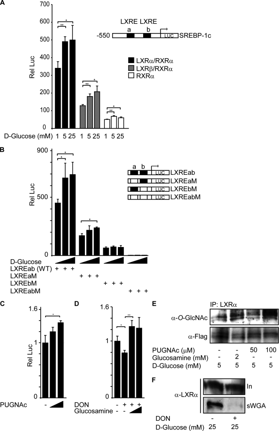
FIGURE 5.
O-GlcNAc regulates LXR transactivation of the SREBP-1c promoter. A, Huh7 cells were transfected with pBP1c550-Luc reporter, Renilla luciferase control plasmid (pRL), and RXRα expression vector together with LXRα (black bars), LXRβ (gray bars), or empty vector pSG5 (white bars). 5 h after transfection, cells were stimulated with different glucose concentrations (1, 5, or 25 mm) for 24 h. A schematic figure of the reporter construct including LXRE elements a and b is shown. B, Huh7 cells were transfected with different pBP1cLXRE-Luc reporter constructs (LXREab, LXREaM, LXREbM, or LXREabM), pRL LXRα, and RXRα expression vectors. 5 h after transfection, cells were stimulated with different glucose concentrations (1, 5, or 25 mm) for 24 h. A schematic figure of the LXRE reporter constructs is shown. C, Huh7 cells were transfected with pBP1cLXREab-Luc reporter, pRL LXRα, and RXRα expression vectors. 5 h after transfection, cells were stimulated with 5 mm glucose in the absence or presence of PUGNAc (50 μm and 100 μm) for 24 h. The luciferase value at 5 mm glucose without stimulation is set as 1. D, Huh7 cells were transfected with pBP1cLXREab-Luc reporter, pRL LXRα, and RXRα expression vectors. 5 h after transfection, cells were stimulated with 25 mm glucose in the absence or presence of DON (5 μm) and glucosamine (0.2 mm and 1 mm) for 24 h. The luciferase value at 25 mm glucose without stimulation is set as 1. All luciferase data are presented as one representative experiment of three or more independent experiments performed in triplicate ± S.D. (error bars). *, p < 0.05; **, p < 0.01. E, LXRα was immunoprecipitated (IP) from FLAG-hLXRα-overexpressing Huh7 cells treated with 5 mm glucose for 24 h in the absence or presence of 2 mm glucosamine or 100 μm PUGNAc. Immunoprecipitated proteins were subjected to SDS-PAGE and blotted with anti-O-GlcNAc (RL2) or anti-FLAG antibodies. F, O-GlcNAc-modified proteins from FLAG-LXRα-overexpressing Huh7 cells cultured for 24 h in 25 mm d-glucose in the absence or presence of DON (5 μm) were absorbed on sWGA-agarose beads. Input (In, 10%) and sWGA pulled-down proteins (sWGA) were subjected to SDS-PAGE and blotted with anti-LXRα antibody.

