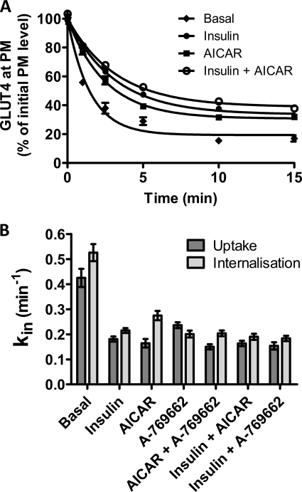
FIGURE 6.
Direct measurements of steady-state GLUT4 internalization. A, L6 myotubes were maintained either in the basal steady state (♦) or in a steady state of stimulation with insulin (●), AICAR (■), or insulin plus AICAR (○). Anti-HA antibody was bound to cell surface GLUT4 at 4 °C; excess antibody was removed by washing, and then the cells were incubated for the indicated times at 37 °C. Surface-bound antibody was then removed, and the remaining antibody was detected with Alexa 488 secondary antibody. Results are the mean ± S.E. from 3 to 5 experiments. In some cases, error bars are smaller than the symbols. B, internalization kin rate constants from steady-state antibody uptake (dark bars) have been compared with the internalization assay measurements (light bars). Standard errors calculated from the confidence intervals (see “Experimental Procedures”) for the fits are shown.

