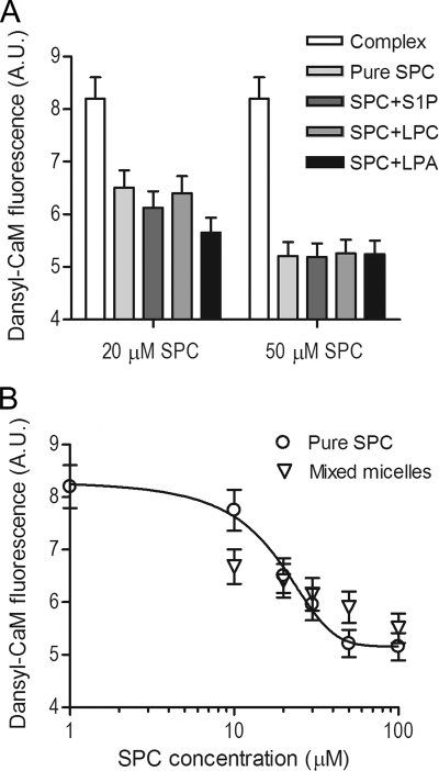
FIGURE 7.
Effect of SPC on the Ca2+-saturated CaM-RyR1 peptide complex in mixed micelles. A, bars depict the fluorescence intensity of 0.2 μm Ca2+-saturated dansyl-CaM with 0.5 μm RyR1 peptide in the absence (white bars) and presence of pure SPC (lightest gray bar) or mixed micelles of various amounts of SPC incorporated into S1P, LPC, or LPA micelles, respectively (darker gray bars). Total lipid concentration was held constant at 100 μm, and mixed micelles contained either 20 or 50 μm SPC. Error bars depict an average experimental error of 5%. B, concentration dependence of the complex dissociating ability of 20% SPC containing LPC micelles (▿) compared with pure SPC (○) taken from Fig. 4E. Ca2+-saturated dansyl-CaM and RyR peptide concentrations were 0.2 and 0.5 μm, respectively. In contrast to panel A, the total lipid concentration varied, and the SPC content was held constant at 20%. Data points represent the mean ± S.E. values of three independent measurements.

