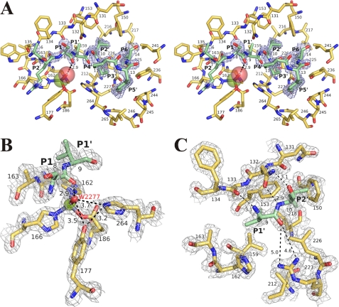
FIGURE 8.
Substrate-binding site of protealysin. A, general stereoview. Omit map contoured at the 3.0σ level in the propeptide region is shown in light blue. B, environment of the catalytic water molecule. C, S1′ and S2′ subsites. In B and C, electron density map contoured at the 1.5σ level is shown in light gray. The catalytic domain is colored yellow orange. The propeptide fragments are colored slate. Propeptide residues (P1′, P2′) are designated after Schechter and Berger (40). The zinc ion is shown as a green sphere; oxygen of HOH-2277, red sphere. The distance between atoms is given in angstroms.

