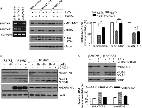
FIGURE 6.
Cathepsin B is involved in ANTXR2-dependent LeTx delivery through autophagic flux. A, HEK293 cells were transfected with control siRNA (si-Scramble) or human (h) ANTXR1- or ANTXR2-specific siRNAs (si-ANTXR1 or si-ANTXR2), and at 42 h post-transfection, portions of the cells were harvested for reverse transcription-PCR (left panel). The remaining cells were incubated in the presence or absence of CA074-Me (100 μm), and treated with LeTx (250 ng/ml LF and 500 ng/ml PA) for 2 h. MEK1 degradation, down-regulation of phospho-ERK (pERK), and LC3-II formation were analyzed by Western blots (middle panel) and MEK1-NT immunoreactivities were analyzed using the NIH-Image program. Data are expressed as mean ± S.D. (n = 3; *, significant, p < 0.05; NS, not significant with p > 0.5). NT, N terminus. B, ANTXR1/R2-deficient CHO cells (R1−/R2−), ANTXR1-reconstituted CHO cells (R1+/R2−), or ANTXR2-reconstituted CHO cells (R1−/R2+) were treated with LeTx (500 ng/ml LF and 1000 ng/ml PA) for the indicated times. MEK1 degradation and LC3-II formation were analyzed by Western blot. Immunoblots for HA or actin were used for transfection or loading control. C, human monocytic THP-1 cells were transfected with control siRNA or siANTXRs, and after 42 h cells were treated with LeTx as above. MEK1 degradation and LC3-II formation were analyzed by Western blot (upper panel), and LC3-II immunoreactivities were analyzed using NHI-Image program (lower panel). Data are expressed as the mean ± S.D. (n = 2).

