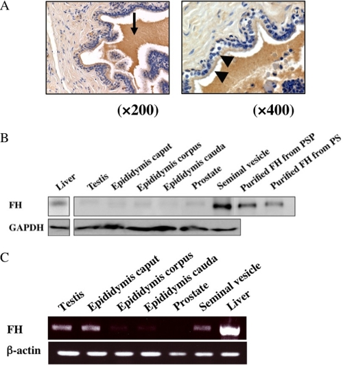
FIGURE 6.
Localization and expression of porcine FH. A, immunohistochemical localization of FH in porcine genital tract tissues. Epithelial cells (arrowhead) and secreted fluid (arrow) in the seminal vesicle were stained, but connective tissue was not stained. Original magnifications: A, left, ×200, and right, ×400. Nuclei were stained with hematoxylin (purple). B, Western blot analysis of various male genital tract tissues. Twenty μg of lysate from each tissue homogenate with RIPA (50 mm Tris-HCl, pH 7.4, 150 mm NaCl 0.1% SDS, 1% sodium cholate, 1% Nonidet P-40, 1 mm EDTA, 0.5 mm phenylmethylsulfonyl fluoride) was immunoblotted with affinity purified anti-FH antibody (upper panel) and anti-GAPDH antibody (lower panel). Lanes from left to right refer to tissue lysates from: liver, testis, epididymis caput, epididymis corpus, epididymis cauda, prostate, and seminal vesicle; and GAPDH was employed as an internal standard. Purified FHs from PSP (eight lane) and PS (far right lane) were used as positive controls. C, expression of FH mRNA in various male genital tract tissues. Twenty-five μg of each tissue was homogenated, and purified RNA was obtained as described under “Experimental Procedures.” RT-PCR products were separated in 1.5% agarose. Lanes from left to right refer to mRNA from: testis, epididymis caput, epididymis corpus, epididymis cauda, prostate, seminal vesicle, and liver. β-Actin was used as an internal control. Liver was used as a positive control.

